Pregnancy scans: what do they show?

Ultrasounds are an exciting milestone on the journey to parenthood. Marianne Falconer outlines what to expect at each scan.
Obstetric ultrasounds are highly anticipated pregnancy milestones. Commonly known as ‘scans’, they provide the parents-to-be with a unique view of the developing foetus while it’s still tucked away in the womb. Often the precious moments spent with the sonographer can be all a woman needs to feel reassured that this pregnancy really is real!
Prospective parents often go to scans with their own expectations, and in the excitement, forget the medical purpose. While most scans unfold perfectly, they can also involve moments of uncertainty and disappointment, so it’s good to be prepared. For this reason, expectant mums often take their partner or a support person along to each appointment.
To help you prepare for these appointments, here’s a guide of what to expect at each ultrasound.
What is ultrasound?
Ultrasound is a non-invasive diagnostic imaging procedure that produces pictures of the inside of the body using high frequency sound waves. The sound waves travel from a small probe called a transducer, through the skin and into the body. They bounce off internal structures like an echo and return to the transducer. The transducer collects these sound waves and sends them to a computer which creates an image. These images are captured in real time and can show the structure and movement of the body’s internal organs. They can also show blood flowing through blood vessels; this is known as Doppler ultrasound.
Ultrasound in pregnancy
During pregnancy, ultrasounds are performed to evaluate a baby’s development. The sonographer will put a clear odourless gel on your stomach and pass the transducer over your skin. This sends out ultrasound waves and picks them up again when they bounce back. A black and white picture of your baby will be shown on a small screen. Most women will have a 12-week scan, a 20-week scan and one growth scan during their ‘singleton’ pregnancy, if it is uncomplicated. However, mothers carrying twins or triplets should be prepared to have more scans.
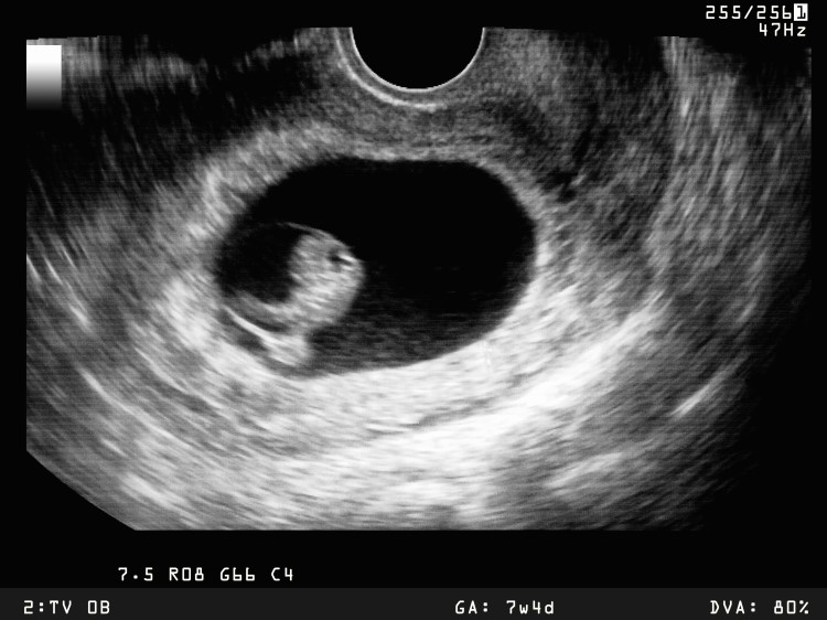

Early pregnancy Scan (7 weeks)
Also known as the ‘dating scan’, this one is non-routine and only offered to mums if needed to assess the dates and viability of the embryo. At seven weeks, the embryo is still tiny (about the size of a blueberry!) so you’ll be asked to drink fluids prior to your appointment to help the sonographer find the cute little button.
Careful measurements taken in the first trimester are even more accurate at predicting an embryo’s age than ultrasounds performed later in pregnancy. As the baby-to-be grows larger, the measurements are less reliable at predicting age because of the varying growth trajectory each baby follows. Highlights at this scan are usually seeing the baby’s heartbeat for the first time (typically visible from 6-7 weeks onwards) and catching a glimpse of your cute blueberry-sized blob, although these observations are not guaranteed.
One of the most awe-inspiring aspects of having an early scan is seeing the incredible growth and development that takes place between now and the routine 12-week scan.
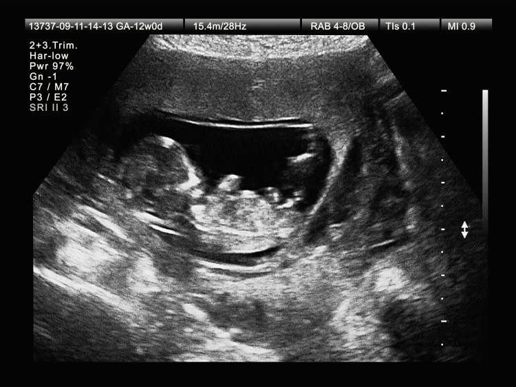

The Chromosomal Assessment Scan
Since the dating scan, the little embryo has developed by leaps and bounds and has transformed into a fully formed foetus –the growth is mind-blowing! At 12 weeks your baby is all of 3 inches long – about the size of a juicy lime! All your baby’s major organs are now developed, so you can expect to see your little one’s vital organs and bone structure, as well as the life-giving placenta.
At this scan you’ll be asked to have a full bladder to aid the sonographer in finding the foetus. The 12-week scan is also called a ‘nuchal translucency scan’ and is a screening test for Down syndrome and other chromosomal abnormalities. The best time to come for a nuchal scan is from 12 weeks until 13.5 weeks, as this increases the scan’s accuracy. The nuchal translucency (or nuchal fold) is a measurement of the skin thickness behind the baby’s neck. This measurement, combined with maternal age and blood test results, gives details of the adjusted chance of having a baby with Down syndrome or other chromosomal abnormality.
The sonographer will also take measurements to ensure your baby is growing appropriately for dates and will check for major abnormalities, however this is limited as the baby is still very small. A more thorough assessment of your baby’s anatomy is done at the 20-week scan.
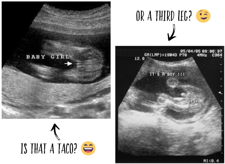

The 20-Week Anatomy Scan
The 20-week scan is often the most anticipated scan as it offers parents-to-be a chance to find out if they’re expecting a girl or a boy. Those in the ‘surprise at birth’ camp will have to look the other way while the sonographer checks the private parts. It’s doubtful whether an untrained eye could spot a ‘third leg’ or a ‘taco’, but its safest to look away anyway, just in case!
By now your baby is about the size of a kumara, and much harder to miss so you won’t have to consume copious amounts of water prior to this scan! The sonographer will:
- Take measurements to check dates and growth.
- Check the position of the placenta.
- Make a thorough examination of the anatomy including:
The spine: Ultrasound is the best diagnostic test for detecting spina bifida.
The heart: At 18 weeks the heart is very small and complex. A detailed scan of the foetal heart will be done at 25-28 weeks in the case of suspected heart abnormality.
The major organs: The presence, appearance and position of all the major organs will be assessed.
The limbs: All four limbs will be fully assessed.
The face: The face will be checked for abnormalities, such as cleft lip.
The head and brain: Brain structures and development will be assessed.
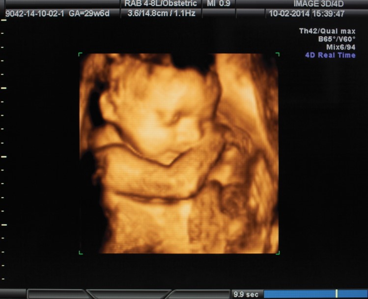

4D scanning offers enthusiastic (slash impatient!) parents a sneak peek of their baby’s face while still in the womb. It’s still not quite the same as the real thing, but the detail is definitely up a level on the traditional 2D scans, and you get a lovely memento for the baby album. Between 26 to 32 weeks is the ideal time for scanning as there is more amniotic fluid and your baby has developed some soft tissue about the face. Be aware that it’s not always possible to get a good image though; baby’s position, the maternal size and the amount of amniotic fluid play a big role in the quality of the 4D scan, and are all out of the sonographer’s control!
What is 4D ultrasound?
4D scanning is the latest ultrasound technology available and can be used to produce a more realistic image of your baby. There is a higher cost associated and only certain clinics offer this new technology. 4D ultrasound is an adjunct to conventional 2D imaging. It uses many conventional 2D images, creates a surface-rendered 3D image, and adds time to the process. On a more serious note, 4D ultrasound is some-times used to enhance visualisation of foetal abnormalities, such as cleft lip, picked up during routine ultrasound scans, so parents can recognise what the doctors are describing.
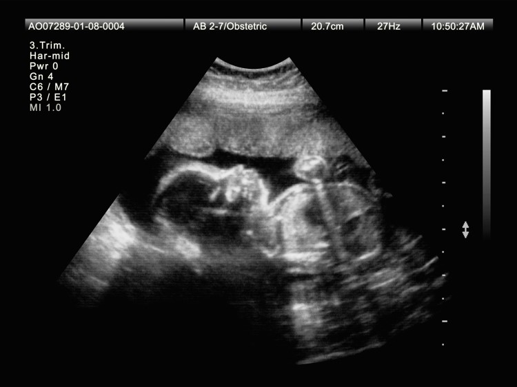

Assessing baby’s growth
Third-trimester growth scans are done to assess your baby’s growth and wellbeing. If there are any concerns, your LMC might request serial growth scans to help indicate when the best time to deliver would be.
Is ultrasound safe?
The use of ultrasound during pregnancy has been commonplace for decades, and it’s generally regarded as the safest and least invasive method for checking the health of a foetus. It’s safe (using sound waves as opposed to the ionising radiation of x-rays), but the focus should not be on scanning for ‘entertainment’ purposes. It’s primarily a medical examination and patients will need a referral from their GP or LMC.
Prenatal diagnosis of abnormalities via ultrasound has had hugely positive effects on the health outcomes of both mothers and their babies, as early detection of some conditions means they can be addressed promptly after birth, or sometimes even in-utero. Improvements in obstetric medicine and ultrasound over the last 30 years have significantly reduced mortality rates in both mothers and babies.
It’s good to remember though, that scans are completely optional and opting out is the mum-to-be’s prerogative. My mum regularly reminds me there were no ultrasounds in her day, and she’s adamant she had less to worry about because of it! However, scans can offer a lovely connection point with your baby-to-be. All the best, parents-to-be, we wish you well on the journey to meeting your little one!
Thanks to Ascot Radiology for supplying the technical information on ultrasound technology and practices.

AS FEATURED IN ISSUE 51 OF OHbaby! MAGAZINE. CHECK OUT OTHER ARTICLES IN THIS ISSUE BELOW


